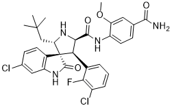
Alpha granule fusion with the platelet membrane causes exposure of P-selectin which by interaction with P-selectin glycoprotein ligand �C 1 Anemarsaponin-BIII mediates the formation of inflammatory platelet-leukocyte complexes. This facilitates a leukocyte influx into the endothelium, thereby presumably assisting in lesion development. Fractional Flow Reserve is an invasive lesion-specific index of myocardial ischemia due to epicardial coronary stenosis. FFR measures a lesion’s ability to cause myocardial ischemia by measuring the pressure gradient across a stenosis during maximum induced hyperemia. Moreover, patients with FFR-positive lesions benefit from revascularization and medical treatment of FFR-positive lesions is inferior to revascularization. We hypothesized that in patients with stable coronary artery disease, the presence of ischemia-causing, flow-limiting coronary lesions, as measured by FFR is associated with altered platelet reactivity. Furthermore, we hypothesized that the presence of hemodynamically significant coronary lesions is associated with altered fractions of platelet-leukocyte complexes. In this observational study we observed no differences in both maximal and cumulative in vitro platelet reactivity between patients with a positive FFR and a negative FFR. However, we did observe a significantly lower percentage PNCs in patients with a positive FFR compared to patients with negative FFR in the clopidogrel treated group, and a trend towards lower percentages of PMCs in patients with a positive FFR compared to a negative FFR in the clopidogrel treated group. Previous clinical studies have shown diverging results with respect to the relationship between platelet  reactivity, plateletleukocyte complexes and coronary luminal obstruction or inducible myocardial ischemia. Increased systemic platelet reactivity was described in patients with documented coronary artery disease immediately after peak exercise, however no relationship with ischemia was found. In critical limb ischemia, increased PMCs and expression of P-selectin was observed, implying an ischemic mechanism of platelet activation. Conversely, other investigators found an inverse relationship between coronary obstruction and platelet reactivity, although all patients investigated had severe single-vessel disease. Others found experimental evidence that platelet reactivity might be reduced by ischemic pre-conditioning, which may point to the possibility of down regulation of platelet reactivity by repeated, short-acting bouts of ischemia as occurs in stable coronary disease. We found no evidence for this. The paradoxical lower percentage of platelet-leukocyte complexes observed in the FFR-positive group may be explained by increased activation of leukocytes due to significantly flow-limiting stenoses and subsequent increased clearance of the complexes from the circulation. Da Costa et al showed that attachment of monocytes to platelets leads to enhanced transmigration of monocytes into the subendothelium. Also, Huo et al showed that the interaction of infused activated platelets with leukocytes resulted in increased adherence to the endothelium. Subsequent transmigration of the complexes led to absence of detectable levels of platelet-leukocyte complexes in a time frame of 3�C4 hours. Acute ischemic events, like myocardial and cerebral infarction cause a strong inflammatory response and tissue damage, and are associated increased levels of peripherally detectable leukocyteplatelet formation during or shortly after the ischemic event, as previously shown. In contrast, inducible ischemia in stable coronary disease implies relatively short, reversible episodes of ischemia without permanent damage, which may transiently increase Isochlorogenic-acid-C leukocyte-platelet complexes.