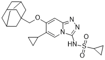
It has been reported that sites of subjectively defined pain, clinically palpated tenderness, tendon thickness and increased colour Doppler signal are Evodiamine anatomically associated, indicating a possible association between pain and neurovascular changes resulting from tendon overuse. However, it must be acknowledged that the colour Doppler signal typically associated with tendinopathy may represent not only angiogenesis, but increased blood flow in vessels which are already present. Angiogenesis may be accompanied by neurogenesis, i.e, nerves may be proliferating along with neovessels in mechanically loaded tendon tissue increasing the level of substance P and other pain-producing substances in tendon. Tenocytes comprise the main cell populationin tendon tissue, and may be defined as scleraxis-expressing fibroblasts residing within the extracellular matrix of the tendon, and playing a key role in tendon development, adaption and the response to mechanical loading. Tenocytes produce a variety of endogenous cytokines and growth factors which exert both autocrine and paracrine effects. Some in vitro studies have shown that repetitive mechanical loading of tendon cells results in an elevated production of soluble factors which are sometimes characterized as inflammatory, catabolic, or anabolic. Several studies have suggested that such changes in gene expression induced by repetitive loading of tenocytes could lead to tendinopathy. In this study, we investigated the expression and activity of angiogenic factors released by cyclically strained, scleraxisexpressing cells derived from human tendon tissue. The prognosis for CRC is based on the formation of distant metastases, not the primary tumor itself. Even with comprehensive research into the biology of cancer progression, the molecular mechanisms involved in the metastatic cascade are not well characterized. The mechanisms of metastasis involve a selective and sequential series of steps, including separation from the primary tumor, invasion through surrounding tissues, entry into the circulatory system, and the establishment and proliferation in a distant location. Two proteins that have been shown to be involved with multiple steps of the metastatic cascade are CD44 and cMET. CD44, a transmembrane glycoprotein that belongs to a family of cell adhesion molecules, is involved with the progression and metastasis of multiple types of cancerand has been associated with a poor prognosis in CRC patients. c-MET is a proto-oncogene that encodes for the receptor tyrosine kinase, also known as hepatocyte growth factor receptor. The only known ligand for c-MET is hepatocyte growth factor ; both c-MET and HGF are upregulated in a number of malignancies and are associated with a poor prognosis and an early predictor of further metastasis. Specifically, c-MET is involved in the regulation of proliferation, motility, invasion and metastasis via its phosphorylation and activation of downstream signaling Forsythin pathways. A comprehensive understanding of the mechanisms that drive CRC metastasis is important for the development of novel approaches to treat this cancer. Therefore, the purpose of our study was to identify the genes that promote liver metastasis in CRC. Here,  we established three highly metastatic CRC cell lines and show that their more aggressive metastatic phenotype is associated with an increase in CD44 expression and activation of c-MET.