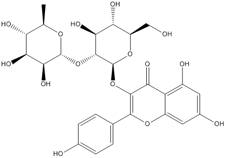
Whereas most of the candidates are functionally similar amongst themselves, in cluster 6 the candidates are all functionally related to the PD-related proteins, with the only exception of YWHAE. A sub-network containing only the candidate proteins that are functionally similar to the PD-related proteins includes 37 proteins in total and 29 candidate Parkin-binding proteins. Twenty-eight candidates fall into four clusters of enriched GO processes that cover the functions of the known PD proteins, including the various known functions of Parkin. Among these processes we have identified several proteins involved in cell death: HSPD1, CASP14, H1F0, DAP3, AIFM1, GSDMA, SET, OPA1, and BAG2. In addition, TOMM70A, DAP3 and OPA1 are involved in mitochondrion organization, and CCT3, CLPX1, HSPD1, PRKCSH and BAG2 are related to protein folding. Of the 29 candidates that are functionally similar to the PD-related proteins, 13 were also identified in a genetic screen as modifiers of Parkin and PINK1 mutant functions in Drosophila, providing further evidence for the biological relevance of the interactions. The diverse functions of the Parkin protein partners reported here are consistent with the functional diversity of the pathogenic processes associated with Parkin-linked parkinsonism. Parkin is localized in the endoplasmic reticulum, the Golgi apparatus, the outer nuclear membrane, synaptic vesicles, and the outer LOUREIRIN-B mitochondrial membrane, and there is a large body of evidence showing that Parkin can interfere with a diverse range of cellular processes and pathways. Like other E3 ubiquitin ligases, it is a component of the ubiquitin proteasome system, a main cellular  pathway that promotes removal of damaged or misfolded proteins, it is involved in signal transduction, protein and membrane trafficking and transcriptional regulation, replication and transcription of mitochondrial DNA, mitophagy, neuroprotection, and apoptosis. Parkin expression has been reported to protect cells against multiple forms of stress, but although the exact mechanism of this prosurvival function remains elusive, accumulating evidence exists that it involves inhibition of programmed cell death. Two recent studies identified Bax and the mitochondrial pro-apoptotic protein ARTS as Parkin substrates that both might contribute to the anti-apoptotic effect of Parkin. In our study, we identified novel associations between Parkin and several proteins involved in cell death processes. An interaction of Parkin with one of them, OPA1, is supported by the observation that inactivation of OPA1, which promotes mitochondrial fusion, rescues the phenotypes of cell death, muscle degeneration, and mitochondrial abnormalities in Parkin and PINK1 mutants in Drosophila. DAP3, another candidate protein involved in cell death, mediates mitochondrial fragmentation, probably reflecting its role in mitochondrial fission. Both proteins might have a role in cell death-associated changes in mitochondrial morphology mediated by Parkin. TOMM70A, which encodes a component of a translocase complex of the outer mitochondrial membrane involved in the import of mitochondrial precursor proteins, has been associated recently to Parkin as it is degraded by the UPS after translocation of Parkin to mitochondria. LRPPRC, which was identified already in a proteomic Diperodon analysis of Parkin interactors, might be involved in mitophagic initiation, maturation, trafficking, and lysosomal clearance through its interaction with the MAP1S protein. Other Parkin-binding proteins, like HSPD1 and CLPX, are involved in protein folding and response to unfolded proteins.