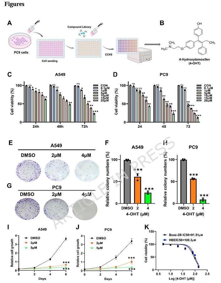
4-Hydroxytamoxifen has spent decades in the shadow of its parent compound, largely catalogued as an estrogen receptor antagonist generated by hepatic metabolism. A recent report in Scientific Reports pulls it into an entirely different spotlight, mapping its effects on MAPK signaling and lipid metabolism in human epithelial cell lines. The investigation began with a high-throughput screen of a human endogenous metabolite library in PC9 cells, where 4-OHT emerged as a potent viability suppressor. Follow-up validation in A549 cells confirmed the initial observation, setting the stage for a mechanistic dissection that links MEK-ERK phosphorylation status to intracellular fatty acid abundance and cellular behavior.
The first layer of evidence came from quantitative proliferation assays. Using a CCK-8 cell counting kit supplied by AbMole, the team measured viability across a concentration gradient spanning 0.1 to 20 μM over 24, 48, and 72 hours. The resulting IC₅₀ curves were steep and time-dependent: A549 cells registered 20.64 μM at 24 hours, dropping to 9.76 μM by 72 hours, while PC9 cells proved even more responsive, moving from 19.41 μM to 6.34 μM over the same interval. What distinguished this profile from generic cytotoxicity was the selectivity window. Non-transformed bronchial epithelial lines HBE and Beas-2B registered IC₅₀ values near 100 μM, indicating that 4-OHT discriminates between transformed and non-transformed phenotypes rather than indiscriminately suppressing all dividing cells. Colony formation assays and crystal violet staining reinforced the CCK-8 data, showing that clonogenic potential and cumulative growth both collapse under sustained 4-OHT exposure in a manner that follows the same dose-response logic.
 Beyond simply slowing division, 4-OHT altered how these cells interact with their surroundings. Wound healing assays showed dramatically reduced closure rates at 5 μM, and transwell migration counts dropped in a dose-dependent fashion. Western blotting offered a molecular explanation for the motility defect: mesenchymal markers vimentin and N-cadherin fell, while epithelial E-cadherin rose. This reversal of epithelial-mesenchymal transition suggests 4-OHT does not merely block proliferation but actively reshapes cellular architecture toward a less motile, more adherent state. Parallel flow cytometry with Annexin V-FITC and propidium iodide revealed a second, distinct mechanism—programmed cell death. The fraction of apoptotic cells climbed steadily with dose, indicating that growth suppression is coupled to activation of cell-intrinsic death programs rather than being a simple consequence of metabolic starvation.
To understand the transcriptional logic driving these phenotypes, the researchers turned to RNA sequencing. Twenty-four hours of 20 μM 4-OHT in A549 cells yielded 701 differentially expressed genes, with 416 upregulated and 285 downregulated. KEGG pathway enrichment placed the MAPK signaling cascade among the most significantly altered pathways. More specifically, a coordinated cluster of fatty acid metabolism genes—including FASN, FADS2, ACAT2, ACSS2, and HMGCR—showed consistent downregulation. Gene set enrichment analysis independently flagged fatty acid degradation and related metabolic processes as significantly altered. These transcriptional shifts pointed toward a metabolic bottleneck rather than a single-gene off-target effect, suggesting that 4-OHT remodels the lipid biosynthetic program at the level of gene expression.
Untargeted metabolomics confirmed the prediction with remarkable clarity. Principal component analysis cleanly separated control and 4-OHT-exposed populations, and orthogonal partial least squares-discriminant analysis reinforced the distinction. Lipid and lipid-like molecules constituted the largest category of depleted metabolites, with numerous fatty acid species showing marked reduction. Direct colorimetric quantification of intracellular free fatty acids showed dose-responsive drops in both A549 and PC9 cells, paralleling the transcriptomic downregulation of synthetic enzymes. The metabolic and transcriptional data converged on a straightforward model: 4-OHT starves cells of lipid building blocks by suppressing de novo fatty acid synthesis at multiple enzymatic steps.
The MAPK connection emerged cleanly from Western blotting. Phosphorylated MEK and phosphorylated ERK both declined sharply after 4-OHT exposure, while total protein levels remained unchanged. This pattern implicates post-translational regulation—specifically, inhibition of kinase activation rather than reduced expression or stability. Since MAPK signaling is known to feed into lipid biosynthetic programs through transcriptional control of rate-limiting enzymes like FASN, the researchers designed rescue experiments to test whether forced reactivation of the pathway could override the metabolic phenotype. Combined exposure with C16-PAF, a selective MAPK activator, partially restored p-MEK and p-ERK levels in both A549 and PC9 cells. More importantly, it rescued free fatty acid content, effectively reversing the lipid depletion caused by 4-OHT alone. Functionally, C16-PAF also restored proliferative capacity in crystal violet assays and migratory behavior in transwell and wound healing experiments. The rescue was partial rather than complete, which is expected given that 4-OHT likely engages multiple parallel mechanisms, but the directionality was unambiguous.
To further tighten the causal inference, the team combined 4-OHT with U0126, a selective MEK1/2 inhibitor. This combination produced deeper suppression of MEK-ERK phosphorylation than either molecule alone, and further accentuated the downregulation of fatty acid metabolism genes. The epistasis is clear: 4-OHT sits upstream of MEK-ERK activation, and the lipid metabolic phenotype is a downstream consequence of reduced pathway flux. The fact that a MEK inhibitor synergizes with 4-OHT rather than duplicating its effect at a ceiling suggests that 4-OHT may impinge on the MAPK axis at a level distinct from MEK itself, or that it simultaneously modulates parallel inputs that converge on ERK.
In vivo validation used a syngeneic C57BL/6J mouse model receiving subcutaneous implantation of LLC cells. Once palpable subcutaneous masses reached approximately 100 mm³, mice received daily intraperitoneal administration of 4-OHT at 40 mg/kg for seven consecutive days. The compound substantially reduced subcutaneous mass volume and weight without triggering body weight loss or hepatorenal histopathology in H&E-stained sections. Immunohistochemical Ki-67 staining fell markedly, while TUNEL positivity rose, mirroring the in vitro proliferation suppression and apoptosis induction. These observations confirm that the MAPK-lipid mechanism operates in an intact physiological setting and is not an artifact of two-dimensional culture.
Several aspects of this study merit emphasis for researchers working at the intersection of signal transduction and metabolism. First, the selectivity for transformed over non-transformed epithelial cells indicates a discrimination window that is not obvious from standard cytotoxicity profiles. Second, the rescue experiments with C16-PAF and U0126 go beyond simple correlation, functionally demonstrating that MEK-ERK phosphorylation status is rate-limiting for both the metabolic and behavioral phenotypes. Third, the integration of transcriptomics and untargeted metabolomics provides a multi-omic anchor for the MAPK-lipid axis that might otherwise be dismissed as a secondary or compensatory effect.
There are limits, of course. The study focused on two related epithelial lines, and the precise transcription factor intermediaries linking MAPK suppression to reduced FASN and HMGCR expression remain uncharacterized. Whether the same mechanism operates in mesenchymal or hematopoietic contexts is an open question. Additionally, the pharmacokinetic behavior of 4-OHT in murine systems was not profiled, so the relationship between the in vitro IC₅₀ and the in vivo dose remains correlative rather than quantitatively linked. Nevertheless, the work establishes 4-OHT as a probe for dissecting the intersection of MAPK signaling and lipid homeostasis, and it positions MEK-ERK-mediated fatty acid synthesis as a central node through which 4-OHT coordinates proliferation, motility, and survival decisions in epithelial cell models.
AbMole Product Integration in This Study
Product: CCK-8 Cell Counting Kit (AbMole, USA)
Application: Quantitative assessment of cell viability and proliferation across human epithelial cell lines to establish dose-response relationships and IC₅₀ values for 4-hydroxytamoxifen.
Experimental Details:
- Cell seeding density: 3,000 cells per well in 96-well plates
- Compound exposure: 4-OHT at concentrations ranging from 0.1 to 20 μM for 24, 48, and 72 hours
- Assay execution: CCK-8 reagent added per manufacturer protocol; absorbance read at 450 nm using a microplate reader
- Analysis: IC₅₀ values calculated via GraphPad Prism 10.0 software
- Cell lines profiled: A549, PC9, HBE, and Beas-2B
Key Findings Enabled by AbMole CCK-8:
- A549 IC₅₀ at 72 h: 9.76 ± 1.9 μM; PC9 IC₅₀ at 72 h: 6.34 ± 2.7 μM
- Non-transformed HBE and Beas-2B bronchial epithelial cells exhibited IC₅₀ values near 100 μM, revealing a roughly tenfold selectivity window
- Dose- and time-dependent viability reduction confirmed across both epithelial lines, with tamoxifen showing comparable IC₅₀ profiles
Meta Description: Mechanistic analysis of 4-OHT-mediated suppression of MEK-ERK phosphorylation and fatty acid metabolism reprogramming, quantified with AbMole CCK-8 assays in human epithelial cell lines.
Target Keywords: 4-hydroxytamoxifen, MAPK signaling, MEK, ERK, lipid metabolism, fatty acid synthesis, FASN, EMT, AbMole, CCK-8, cell viability